Diabetes affects blood vessels throughout the body. The eye contains some of the most delicate vessels of all. Inside the retina, these capillaries regulate oxygen and nutrients for vision. When blood sugar stays high, the vessel walls thicken or leak. Fluid can build behind the retina, blurring the image. Over time, the eye loses its ability to process light correctly. This process starts slowly but causes permanent damage without early intervention.
In many cases, damage begins without pain or visible symptoms
Most patients don’t notice changes in the early stages. Vision may stay sharp for months. But microscopic swelling may already be occurring. This is why diabetic eye exams are not optional. Waiting for symptoms delays treatment. The condition progresses silently, hidden behind clear vision. Many discover it only when irreversible damage has occurred. By then, treatment options are limited and outcomes are less predictable.
Diabetic retinopathy is the most common cause of blindness in working-age adults worldwide
This condition involves blood vessels breaking down in the retina. In mild forms, small leaks appear. As it advances, the body forms new vessels to compensate. But these new vessels are fragile. They can bleed into the eye, causing dark spots or total vision loss. Scar tissue may pull on the retina, risking detachment. In advanced stages, even surgery may not fully restore function.
Macular edema occurs when fluid builds near the center of the retina
The macula controls our sharpest central vision. Reading, driving, and recognizing faces depend on it. When edema sets in, the macula swells. Vision becomes wavy or cloudy. Bright light may cause discomfort. Colors lose intensity. This complication can develop at any stage of diabetic retinopathy. It’s often the main reason patients seek help. Unfortunately, by that point, treatment becomes more complex.
New vessels can bleed into the vitreous, clouding the eye from within
When new vessels form during proliferative retinopathy, they tend to rupture easily. Blood fills the vitreous gel. Patients report seeing floaters or red shadows. In some cases, vision drops suddenly and dramatically. This condition may require laser treatment or vitrectomy. The blood itself is harmless but blocks light. If not cleared, it can cause scarring that damages the retina further.
Some patients develop neovascular glaucoma as a secondary consequence of uncontrolled retinopathy
In this condition, new vessels grow over the iris and drainage channels. Pressure builds inside the eye. Unlike typical glaucoma, this form resists common treatments. Pain and redness may follow. If pressure remains high, optic nerve damage can occur. Loss of peripheral vision becomes permanent. This condition links directly to late-stage diabetic changes in the retina.
The lens becomes cloudy faster in diabetic patients, even at younger ages
Cataracts often appear earlier in people with diabetes. High sugar levels alter the lens structure. Water enters the lens, distorting vision. Blurriness and glare from lights become frequent complaints. Cataracts in diabetics tend to progress faster. Surgery remains effective, but outcomes depend on retinal health. If retinopathy coexists, vision may not fully improve after lens replacement.
Fluctuating blood sugar can cause temporary shifts in focusing power
Some patients notice their vision changing week to week. Eyeglass prescriptions seem wrong suddenly. These changes reflect swelling in the lens from sugar imbalances. They may not signal permanent damage but interfere with daily life. Stable glucose levels are essential before prescribing glasses. Optometrists often wait weeks to test vision after big sugar swings.
Diabetes affects the nerves that control pupil reaction and tear production
Some diabetics develop autonomic neuropathy involving the eyes. Pupils may react slowly to light. Night driving becomes harder. Dryness increases due to reduced tear secretion. This leads to irritation, redness, and risk of infection. Artificial tears can relieve discomfort, but the underlying nerve issue remains. These changes are less visible but still impact comfort and safety.
Pregnancy with diabetes increases the risk of rapid eye deterioration
Hormonal shifts in pregnancy add pressure to already strained vessels. Gestational diabetes may trigger changes too. Women with existing diabetes should undergo detailed eye checks early in pregnancy. Vision changes may happen suddenly. Controlling glucose tightly during this time is essential. Delays may risk both mother and fetus.
Laser photocoagulation helps seal leaking vessels and slow the progression of retinopathy
In this procedure, a laser targets damaged vessels. Heat seals them shut. It reduces swelling and halts further growth. Vision may not improve but deterioration slows. Some peripheral vision is lost, especially after extensive laser treatment. But central vision is often preserved. It’s a trade-off many accept to protect overall sight.
Injections into the eye reduce swelling and block abnormal vessel growth
Anti-VEGF drugs are injected directly into the vitreous. These medications block signals that promote new vessels. They also shrink existing abnormal ones. Treatments are repeated every month at first. Some patients stabilize and need fewer injections later. This approach is effective for macular edema and early proliferative retinopathy. It doesn’t reverse damage but slows it down significantly.
Vitrectomy removes blood and scar tissue from the back of the eye
This surgical procedure becomes necessary in advanced cases. The surgeon removes the vitreous gel. Scar tissue is dissected. The goal is to restore light flow to the retina. Recovery takes weeks. Success depends on the retina’s condition. Some patients regain useful vision. Others see limited improvement. It’s often the final option after laser and injections.
Eye complications develop faster in those with longer diabetes duration
Time plays a key role. The longer someone lives with diabetes, the higher the risk. Retinopathy may not appear for the first five years. But by year fifteen, it’s common. Type 1 and type 2 carry similar long-term risks. However, early diagnosis and strict glucose control delay progression. Routine checks make the difference.
Blood pressure and cholesterol influence diabetic eye complications significantly
It’s not just sugar. High blood pressure strains eye vessels too. Cholesterol clogs them. When combined, these factors speed up retinopathy. That’s why eye health requires full-body care. A normal A1C isn’t enough if pressure stays high. Managing all risks together gives the best chance for long-term vision.
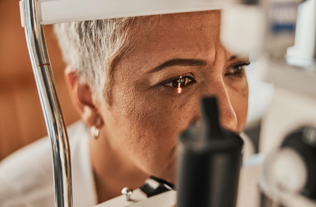
Annual dilated eye exams are essential even when vision seems normal
These exams allow detailed inspection of the retina. Dilation reveals tiny leaks or abnormal vessels. Photos and scans track changes over time. Early signs may appear before vision changes. Detecting them allows earlier treatment. Patients who skip these exams risk late discovery. Prevention depends on showing up regularly.
Some technologies now allow home-based monitoring for retinal changes
New tools let patients photograph their eyes at home. Apps can detect swelling patterns. These innovations don’t replace clinical exams but offer extra safety. For those in remote areas, they add value. AI-assisted tools analyze patterns missed by the human eye. But interpretation still requires specialists.
Even with modern treatments, permanent vision loss still occurs in many patients
Despite lasers, injections, and surgeries, outcomes are not guaranteed. Some damage can’t be undone. The retina does not regenerate well. Scarred areas don’t regain function. The goal shifts to preventing further loss. This is why early action matters more than any single treatment. Catching changes early is the best strategy.
Sight remains vulnerable without lifelong commitment to glucose and pressure control
Control must continue forever. One good year doesn’t erase ten bad ones. Lapses lead to relapse. Eye complications return fast after poor control. There’s no finish line—only ongoing care. Patients who stay consistent usually retain usable vision. Others face slow decline. The difference lies in daily decisions.