High blood pressure can damage blood vessels in the eyes, leading to a condition called hypertensive retinopathy. These vessels are delicate and respond quickly to changes in systemic pressure. When blood pressure remains elevated, the retinal vessels thicken, narrow, or leak fluid. Over time, this affects how the retina functions, possibly distorting vision or causing blind spots. Retinopathy severity often reflects how long hypertension has been uncontrolled. The condition may go unnoticed until damage is significant. Routine eye exams can reveal early signs before symptoms appear. Catching changes early offers a chance to protect remaining vision.
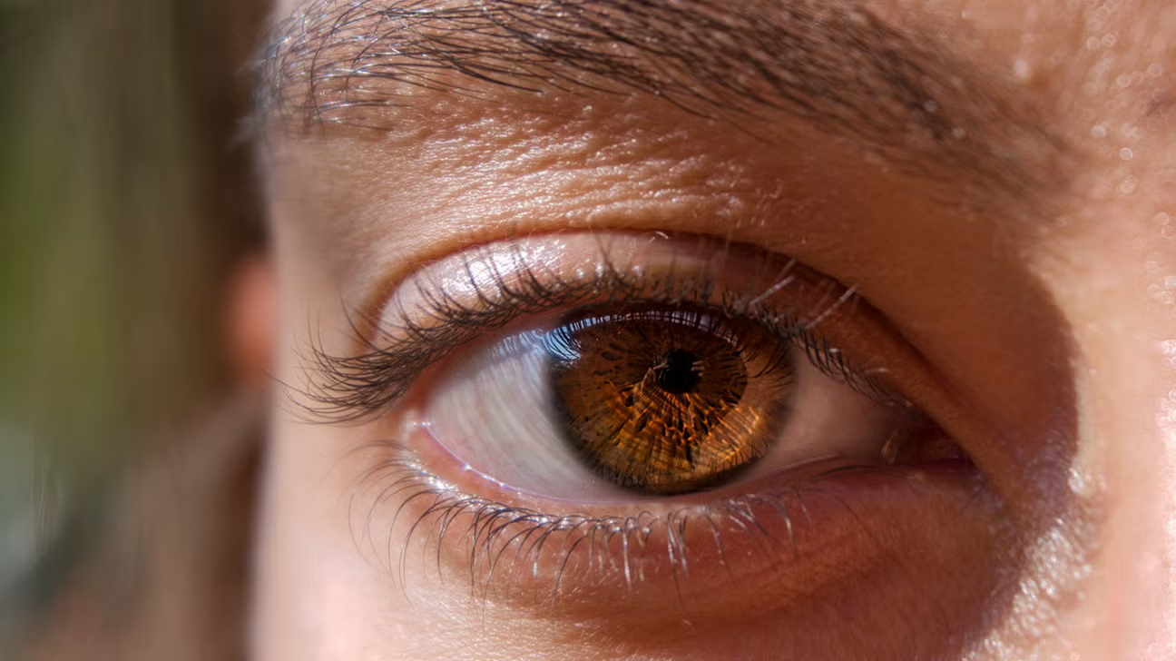
Retinal arteries may harden and become less flexible, reducing blood flow to light-sensitive tissues in the eye
Retinal arteries may harden and become less flexible, reducing blood flow to light-sensitive tissues in the eye. This stiffness limits oxygen delivery where it’s most needed. The retina depends on a steady supply of nutrients to process visual information. When blood flow drops, retinal cells struggle and begin to fail. The damage can be permanent if left unchecked. Vision may blur, particularly during blood pressure spikes or stress. These episodes are often temporary at first but grow longer with time. Arterial narrowing is detectable through dilated eye exams and fundus imaging by trained professionals.
In some cases, blood vessels leak fluid or blood, creating swelling in the central retina, or macula
In some cases, blood vessels leak fluid or blood, creating swelling in the central retina, or macula. This area controls sharp vision and detail perception. When it swells, tasks like reading or recognizing faces become difficult. The swelling is called macular edema and often worsens silently. Patients may first notice distortion or gray patches in their field of view. If untreated, the swelling can damage nerve layers permanently. Imaging tests such as OCT help identify fluid early. Treatment focuses on controlling blood pressure and sometimes includes eye injections to reduce inflammation and leakage.
Sudden spikes in blood pressure may lead to optic nerve damage, threatening central vision over time
Sudden spikes in blood pressure may lead to optic nerve damage, threatening central vision over time. The optic nerve transmits signals from the retina to the brain. When blood pressure rises rapidly, circulation to this nerve can be compromised. A condition called ischemic optic neuropathy may occur, often without warning. Patients describe sudden vision dimming or dark areas in one eye. The nerve’s sensitivity makes recovery difficult once damage sets in. Management involves systemic pressure control and sometimes neuroprotective strategies. Identifying risk factors like sleep apnea or vascular disease can lower the chance of nerve injury.
Hypertensive retinopathy progresses silently, meaning damage often occurs before patients notice visual changes
Hypertensive retinopathy progresses silently, meaning damage often occurs before patients notice visual changes. Early stages may show no symptoms at all. Regular eye exams become essential in high-risk individuals. Subtle signs like vessel narrowing or retinal hemorrhages indicate worsening disease. By the time symptoms develop, damage is usually advanced. Vision loss from hypertension is preventable but often overlooked. Ophthalmologists use grading systems to assess severity and progression. These grades help guide treatment urgency and medical follow-up frequency. Delay in diagnosis limits options for preserving function and avoiding irreversible changes.
Vision loss caused by chronic high blood pressure may become permanent if left unmanaged for too long
Vision loss caused by chronic high blood pressure may become permanent if left unmanaged for too long. The cumulative effects on vessels, nerves, and retinal tissue become harder to reverse. Some patients recover partially after blood pressure stabilizes. However, once retinal cells die or nerves atrophy, function cannot return. Early detection and pressure control are the best defenses against progression. Eye damage rarely improves without addressing systemic health. Medications, diet changes, and exercise protect more than just the heart. They help preserve eye health across decades. Regular communication between eye doctors and primary care improves outcomes.
Other hypertensive complications, like stroke, may also affect vision by disrupting brain signals to the eyes
Other hypertensive complications, like stroke, may also affect vision by disrupting brain signals to the eyes. The visual pathway includes several brain regions beyond the eyeball itself. Stroke in areas like the occipital lobe can cause field cuts or complete blindness. Even small clots or bleeds can interrupt signal transmission. These events often accompany severe or untreated hypertension. Recovery depends on location and extent of brain involvement. Rehabilitation may help retrain visual processing, but outcomes vary. Managing blood pressure lowers the chance of such neurological vision loss. Prevention remains more effective than any form of recovery therapy.
People with diabetes and high blood pressure face even greater risk for severe vision complications
People with diabetes and high blood pressure face even greater risk for severe vision complications. These conditions accelerate vascular damage and inflammation inside the retina. Diabetic retinopathy and hypertensive retinopathy may occur together, worsening each other’s effects. Blood vessels leak, swell, or close off entirely. Combined, they raise the risk of retinal detachment or severe macular damage. Tight control of both glucose and pressure becomes essential. Endocrinologists and ophthalmologists often coordinate care in such cases. Ignoring either factor can speed up vision loss and reduce treatment effectiveness. Patients need aggressive management, not just routine checkups.
Hypertensive changes in the eye are visible through a dilated eye exam with specialized imaging techniques
Hypertensive changes in the eye are visible through a dilated eye exam with specialized imaging techniques. Fundus photography captures vessel condition, bleeding, and swelling signs. Optical coherence tomography scans retinal layers in detail, detecting subtle fluid buildup. Fluorescein angiography can reveal hidden leaks or circulation problems. These tools guide decisions about monitoring or urgent intervention. They also document disease progression for long-term tracking. Without such tools, early-stage hypertensive retinopathy often goes unnoticed. Annual eye exams help detect vascular stress before symptoms occur. This monitoring allows early changes in blood pressure treatment to preserve vision.
Preventing hypertensive eye damage requires consistent blood pressure control, healthy habits, and regular eye screenings
Preventing hypertensive eye damage requires consistent blood pressure control, healthy habits, and regular eye screenings. Managing lifestyle plays a key role in long-term outcomes. Salt reduction, stress management, and exercise all support stable blood flow. Medication adherence ensures pressure stays within target range daily. Smoking and excessive alcohol worsen vascular reactivity and should be addressed. Eye doctors can catch vessel changes early if patients attend appointments regularly. Pressure numbers alone don’t reflect total risk. Comprehensive care means watching both numbers and organs over time.